Volume 1 Issue 1 2020
Click Here to Download Full IssueEDITORIAL
2020, 1(1): 1–2. doi:https://doi.org/10.3877/cma.j.issn.2096-112X.2020.01.001
REVIEW
2020, 1(1): 3–17. doi:https://doi.org/10.3877/cma.j.issn.2096-112X.2020.01.002
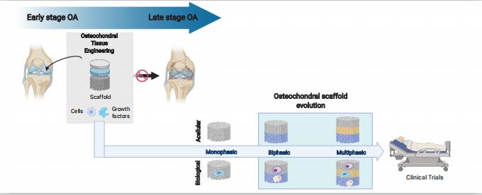
Osteoarthritis is a degenerative joint disease, typified by the loss in the quality of cartilage and bone at the interface of a synovial joint, resulting in pain, stiffness and reduced mobility. The current surgical treatment for advanced stages of the disease is joint replacement, where the non-surgical therapeutic options or less invasive surgical treatments are no longer effective. These are major surgical procedures which have a substantial impact on patients’ quality of life and lifetime risk of requiring revision surgery. Treatments using regenerative methods such as tissue engineering methods have been established and are promising for the early treatment of cartilage degeneration in osteoarthritis joints. In this approach, 3-dimensional scaffolds (with or without cells) are employed to provide support for tissue growth. However, none of the currently available tissue engineering and regenerative medicine products promotes satisfactory durable regeneration of large cartilage defects. Herein, we discuss the current regenerative treatment options for cartilage and osteochondral (cartilage and underlying subchondral bone) defects in the articulating joints. We further identify the main hurdles in osteochondral scaffold development for achieving satisfactory and durable regeneration of osteochondral tissues. The evolution of the osteochondral scaffolds - from monophasic to multiphasic constructs - is overviewed and the osteochondral scaffolds that have progressed to clinical trials are examined with respect to their clinical performances and their potential impact on the clinical practices. Development of an osteochondral scaffold which bridges the gap between small defect treatment and joint replacement is still a grand challenge. Such scaffold could be used for early treatment of cartilage and osteochondral defects at early stage of osteoarthritis and could either negate or delay the need for joint replacements.
REVIEW
2020, 1(1): 18–32. doi:https://doi.org/10.3877/cma.j.issn.2096-112X.2020.01.003
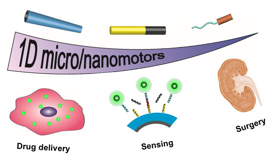
The rapid development of artificial micro/nanomachines brings promising strategies to overcome challenges in biomedicine, including delivery, sensing and surgery. One-dimensional (1D) micro/nanomotors are one of the most attractive micro/nanomachines due to their high specific surface area, powerful impetus and weak rotation diffusion. In this review, different propulsion mechanisms and motion control strategies of 1D micro/nanomotors are summarized, and recent efforts towards their fabrication methods and biomedical applications are discussed. We envision the multidisciplinary research efforts in the field of 1D micro/nanomotors will pave their way to practical applications in bioimaging and biomedicine.
REVIEW
2020, 1(1): 33–45. doi:https://doi.org/10.3877/cma.j.issn.2096-112X.2020.01.004
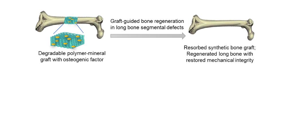
Recent developments in synthetic bone grafting materials and adjuvant therapeutic agents have opened the door to the regenerative reconstruction of critical-size long bone segmental defects resulting from trauma, osteoporotic fractures or tumour resections. Polymeric scaffolds with controlled macroporosities, degradability, useful surgical handling characteristics, and the ability to deliver biotherapeutics to promote new bone ingrowth have been developed for this challenging orthopaedic application. This review highlights major classes of degradable synthetic polymers and their biomineral composites, including conventional and amphiphilic polyesters, polyanhydrides, polycarbonates, and polyethylene glycol-based hydrogels, that have been explored for the regenerative reconstruction of critical-size long bone segmental defects over the past two decades. The pros and cons of these synthetic scaffold materials are presented in the context of enabling or impeding the functional (mechanical and radiographic) repair of a long bone segmental defect, with the long bone regeneration outcomes compared with healthy long bone controls or results achieved with current grafting standards.
REVIEW
2020, 1(1): 46–57. doi:https://doi.org/10.3877/cma.j.issn.2096-112X.2020.01.005
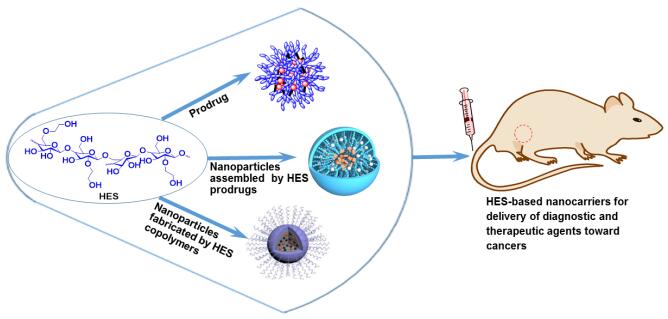
Many types of drugs and agents used for cancer diagnosis and therapy often have low bioavailability and insufficient efficacy, as well as causing various side effects due to their nonspecific delivery. Nanocarriers with purposely-designed compositions and structures have shown varying degrees of abilities to deliver these compounds towards cancers in passive or active manners. Despite the availability of a variety of materials for the construction of nanocarriers, natural polymers with good biocompatibility and biodegradability are preferable for such usage because of their high in vivo safety as well as easy removal of degradation products. Among the natural polymers intended for building nanocarriers, hydroxyethyl starch and its derivatives have gained tremendous attention in the field of drug delivery in the form of nanomedicines over the last decade. There is growing optimism that ever more hydroxyethyl starch-based nanomedicines will be a significant addition to the armoury currently used for cancer diagnosis and therapy.
REVIEW
2020, 1(1): 58–68. doi:https://doi.org/10.3877/cma.j.issn.2096-112X.2020.01.006
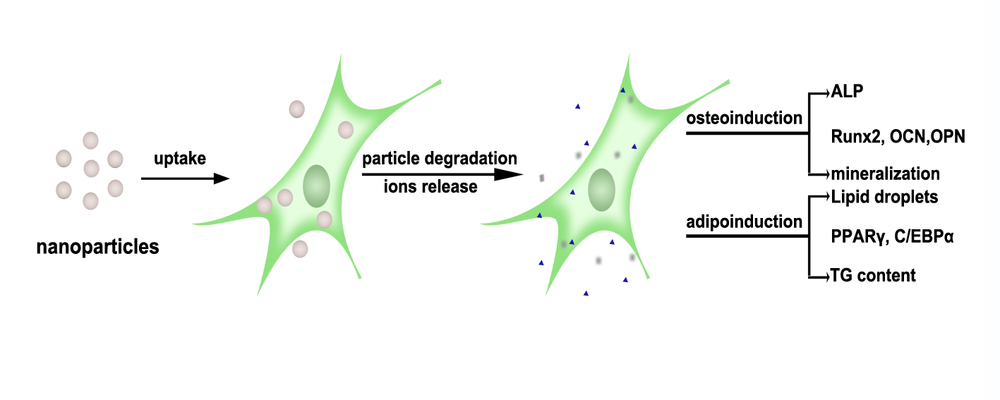
Over the past decades, advancements in nanoscience and nanotechnology have resulted in numerous nanomedicine platforms. Various nanoparticles, which exhibit many unique properties, play increasingly important roles in the field of biomedicine to realize the potential of nanomedicine. Due to the capacity of self-renewal and multilineage mesenchymal differentiation, mesenchymal stem cells (MSCs) have been widely used in the area of regenerative medicine and in clinical applications due to their potential to differentiate into various lineages. There are several factors that impact the differentiation of MSCs into different lineages. Many types of biomaterials such as polymers, ceramics, and metals are commonly applied in tissue engineering and regenerative therapies, and they are continuously refined over time. In recent years, along with the rapid development of nanotechnology and nanomedicine, nanoparticles have been playing more and more important roles in the fields of biomedicine and bioengineering. The combined use of nanoparticles and MSCs in biomedicine requires greater knowledge of the effects of nanoparticles on MSCs. This review focuses on the effects of four inorganic or metallic nanoparticles (hydroxyapatite, silica, silver, and calcium carbonate), which are widely used as biomaterials, on the osteogenic and adipogenic differentiation of MSCs. In this review, the cytotoxicity of these four nanoparticles, their effects on osteogenic/adipogenic differentiation of MSCs and the signalling pathways or transcription factors involved are summarized. In addition, the chemical composition, size, shape, surface area, surface charge and surface chemistry of nanoparticles, have been reported to impact cellular behaviours. In this review, we particularly emphasize the influence of their size on cellular responses. We envision our review will provide a theoretical basis for the combined application of MSCs and nanoparticles in biomedicine.
RESEARCH ARTICLE
2020, 1(1): 69–81. doi:https://doi.org/10.3877/cma.j.issn.2096-112X.2020.01.007
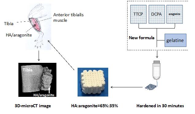
A self-hardening three-dimensional (3D)-porous composite bone graft consisting of 65 wt% hydroxyapatite (HA) and 35 wt% aragonite was fabricated using a 3D-Bioplotter®. New tetracalcium phosphate and dicalcium phosphate anhydrous/aragonite/gelatine paste formulae were developed to overcome the phase separation of the liquid and solid components. The mechanical properties, porosity, height and width stability of the end products were optimised through a systematic analysis of the fabrication processing parameters including printing pressure, printing speed and distance between strands. The resulting 3D-printed bone graft was confirmed to be a mixture of HA and aragonite by X-ray diffraction, Fourier transform infrared spectroscopy and energy dispersive X-ray spectroscopy. The compression strength of HA/aragonite was between 0.56 and 2.49 MPa. Cytotoxicity was assessed using the 3-(4,5-dimethylthiazol-2-yl)-2,5-diphenyltetrazolium bromide (MTT) assay in vitro. The osteogenicity of HA/aragonite was evaluated in vitro by alkaline phosphatase assay using human umbilical cord matrix mesenchymal stem cells, and in vivo by juxtapositional implantation between the tibia and the anterior tibialis muscle in rats. The results showed that the scaffold was not toxic and supported osteogenic differentiation in vitro. HA/aragonite stimulated new bone formation that bridged host bone and intramuscular implants in vivo. We conclude that HA/aragonite is a biodegradable and conductive bone formation biomaterial that stimulates bone regeneration. Since this material is formed near 37°C, it will have great potential for incorporating bioactive molecules to suit personalised application; however, further study of its biodegradation and osteogenic capacity is warranted. The study was approved by the Animal Ethical Committee at Tongji Medical School, Huazhong University of Science and Technology (IACUC No. 738) on October 1, 2017.
RESEARCH ARTICLE
2020, 1(1): 82–88. doi:https://doi.org/10.3877/cma.j.issn.2096-112X.2020.01.008
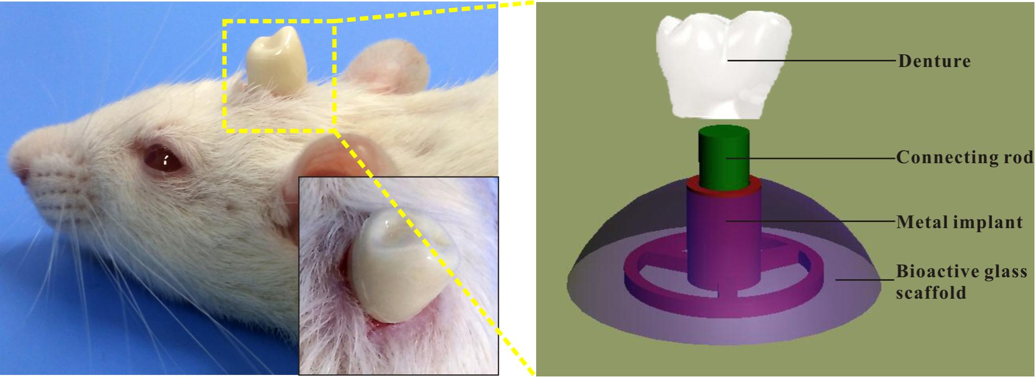
Alveolar ridge atrophy brings great challenges for endosteal implantation due to the lack of adequate vertical bone mass to hold the implants. To overcome this limitation, we developed a novel dental implant design: sub-scaffold dental implant system (SDIS), which is composed of a metal implant and a micro-nano bioactive glass scaffold. This implant system can be directly implanted under mucous membranes without adding any biomolecules or destroying the alveolar ridge. To evaluate the performance of the novel implant system in vivo, SDISs were implanted into the sub-epicranial aponeurosis space of Sprague-Dawley rats. After 6 weeks, the SDIS and surrounding tissues were collected and analysed by micro-CT, scanning electron microscopy and histology. Our results showed that SDISs implanted into the sub-epicranial aponeurosis had integrated with the skull without any mobility and could stably support a denture. Moreover, this design achieved alveolar ridge augmentation, as active osteogenesis could be observed outside the cortical bone. Considering that the microenvironment of the sub-epicranial aponeurosis space is similar to that of the alveolar ridge, SDISs have great potential for clinical applications in the treatment of atrophic alveolar ridges. The study was approved by the Animal Care Committee of Guangdong Pharmaceutical University (approval No. 2017370).
RESEARCH ARTICLE
2020, 1(1): 89–98. doi:https://doi.org/10.3877/cma.j.issn.2096-112X.2020.01.009
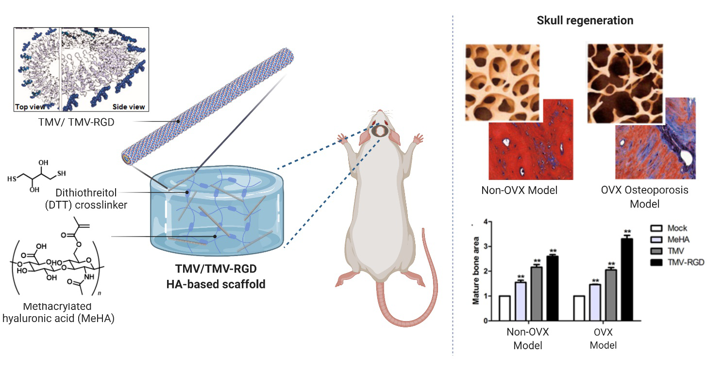
Tobacco mosaic virus (TMV) has been studied as a multi-functional agent for bone tissue engineering. An osteo-inductive effect of wild-type TMV has been reported, as it can significantly enhance the bone differentiation potential of bone marrow stromal cells both on a two-dimensional substrate and in a three-dimensional (3D) hydrogel system. A TMV mutant (TMV-RGD1) was created which featured the adhesion peptide arginyl-glycyl-aspartic acid (RGD), the most common peptide motif responsible for cell adhesion to the extracellular matrix, on the surface of the virus particle to enhance the bio-functionality of the scaffold material. We hypothesised that the incorporation of either wild-type TMV or TMV-RGD1 in the 3D hydrogel scaffold would induce bone healing in critical size defects of the cranial segmental bone. We have previously tested the virus-functionalised scaffolds, in vitro, with a hyaluronic acid-based system as an in-situ hydrogel platform for 3D cell encapsulation, culture, and differentiation. The results of these experiments suggested the potential of the virus-functionalised hydrogel to promote in vitro stem cell differentiation. The hydrogel-forming system we employed was shown to be safe and biocompatible in vivo. Here, we further explored the physiological responses regarding bone regeneration of a calvarial defect in both normal and osteoporotic ovariectomized rat models. Our results, based on histological analysis in both animal models, suggested that both wild-type TMV and TMV-RGD1 functionalised hydrogels could accelerate bone regeneration, without systemic toxicity, evaluated by blood counts. New bone formation was intensified by the incorporation of the RGD-mutant viral particles. This finding increased the potential for use of the rod-shaped plant virus as a platform for the addition of powerful biofunctionality for tissue engineering applications. This study was approved by the Ethics Committee on Animal Use of the Zhenjiang Affiliated First People’s Hospital affiliated to Jiangsu University.