News and Announcements
Volume 3 Issue 3 2022
Order by
Latest time
EDITORIAL
A milestone towards a successful scientific journal: celebrating the inclusion of Biomaterials Translational by PubMed
Qian Wang
3 Download 1125 Views
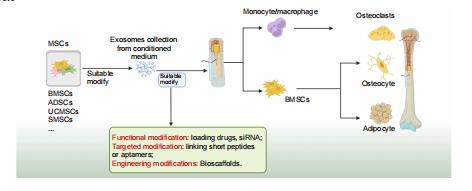
REVIEW
Mesenchymal stem cell–derived extracellular vesicles: a possible therapeutic strategy for orthopaedic diseases: a narrative review
Zhao–Lin Zeng,
Hui Xie
14 Download 2294 Views
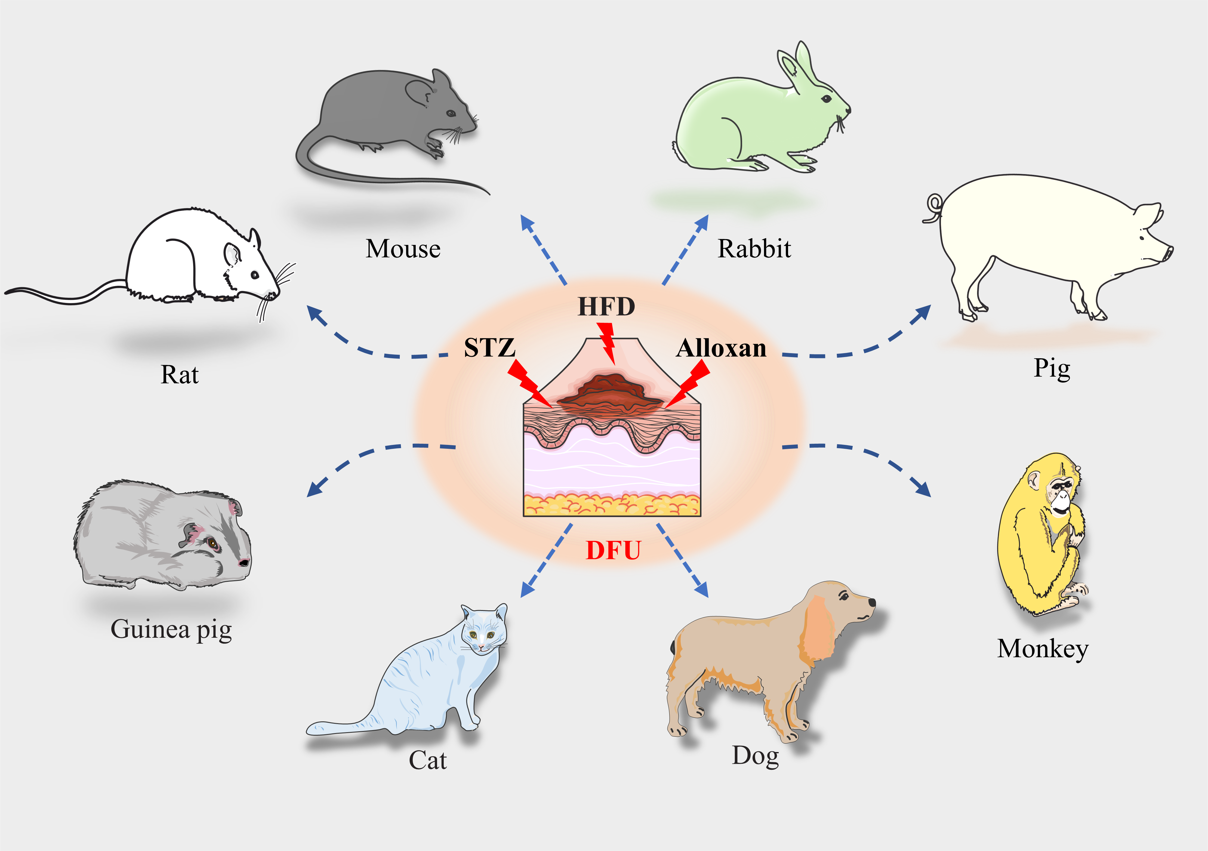
REVIEW
Advances and perspective on animal models and hydrogel biomaterials for diabetic wound healing
Yiqiang Hu,
Yuan Xiong,
Ranyang Tao,
Hang Xue, ... Guohui Liu
93 Download 7477 Views
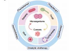
REVIEW
Engineered microorganism–based delivery systems for targeted cancer therapy: a narrative review
Xin Huang,
Haoyu Guo,
Lutong Wang,
Zengwu Shao
18 Download 1908 Views
RESEARCH ARTICLE
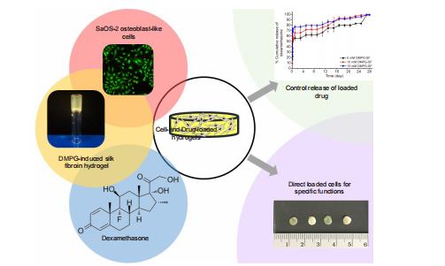
Osteogenic differentiation of encapsulated cells in dexamethasone–loaded phospholipid–induced silk fibroin hydrogels
Chavee Laomeephol,
Helena Ferreira,
Sorada Kanokpanont,
Jittima Amie Luckanagul, ... Siriporn Damrongsakkul
16 Download 1416 Views
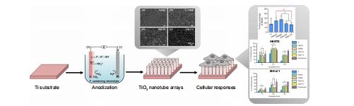
RESEARCH ARTICLE
Cellular responses to nanoscale substrate topography of TiO2 nanotube arrays: cell morphology and adhesion
Monchupa Kingsak,
Panita Maturavongsadit,
Hong Jiang,
Qian Wang
18 Download 1567 Views