
Human–Artificial intelligence dialogue: Beyond the language barrier
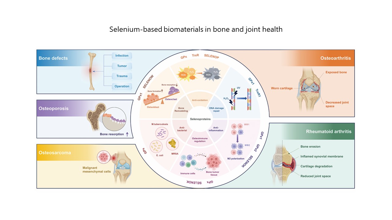
Advances in selenium research for bone and joint-related diseases: from pathophysiological mechanisms to therapeutic implications of selenium-based biomaterials
Functionalization and rehabilitation applications of sodium alginate-based hydrogels

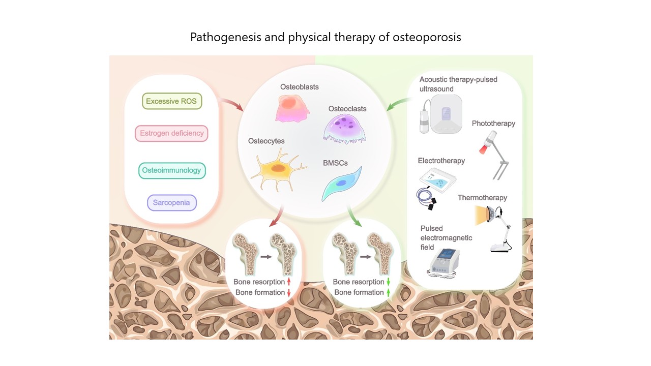
Advancements in physical therapy for osteoporosis treatment
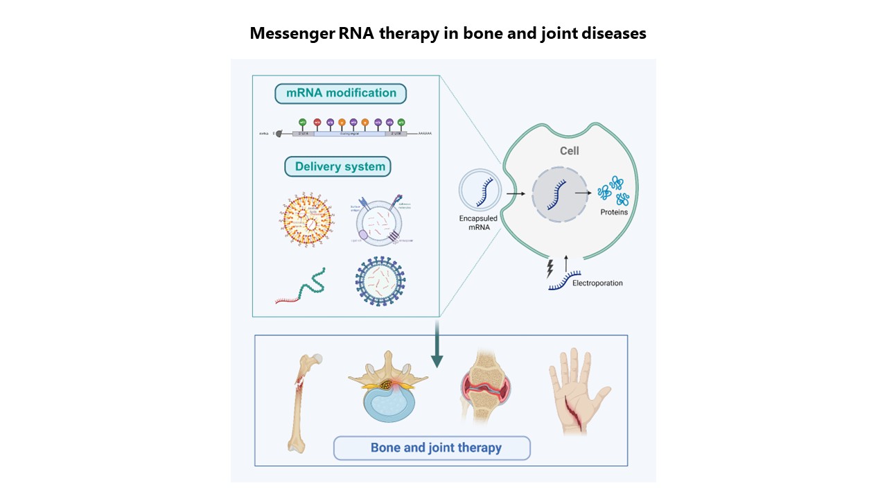
Messenger RNA therapy in bone and joint diseases: Rationale, delivery systems, and applications
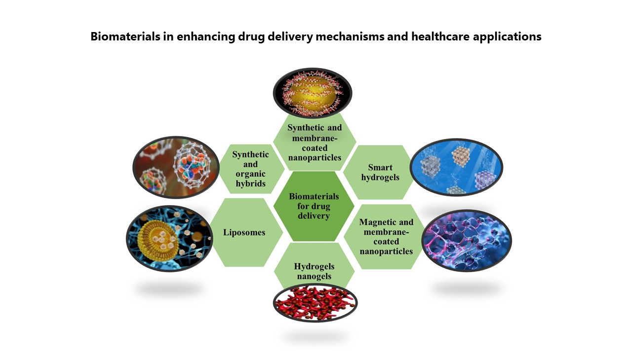
Transforming therapeutics through biomaterials: A comprehensive insight into biomaterials’ role in effective drug delivery and healthcare advancement
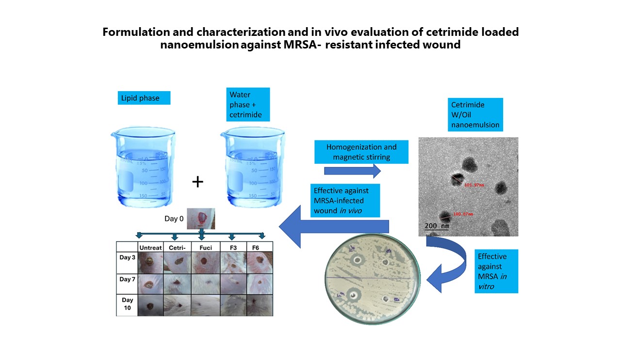
Topical triple-strategy nanoemulsion formulations of cetrimide for treating methicillin-resistant Staphylococcus aureus-infected skin wounds
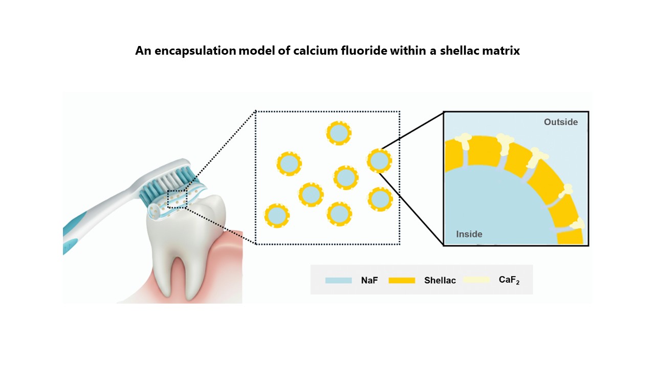
Shellac-based encapsulation model incorporating calcium fluoride for oral care applications
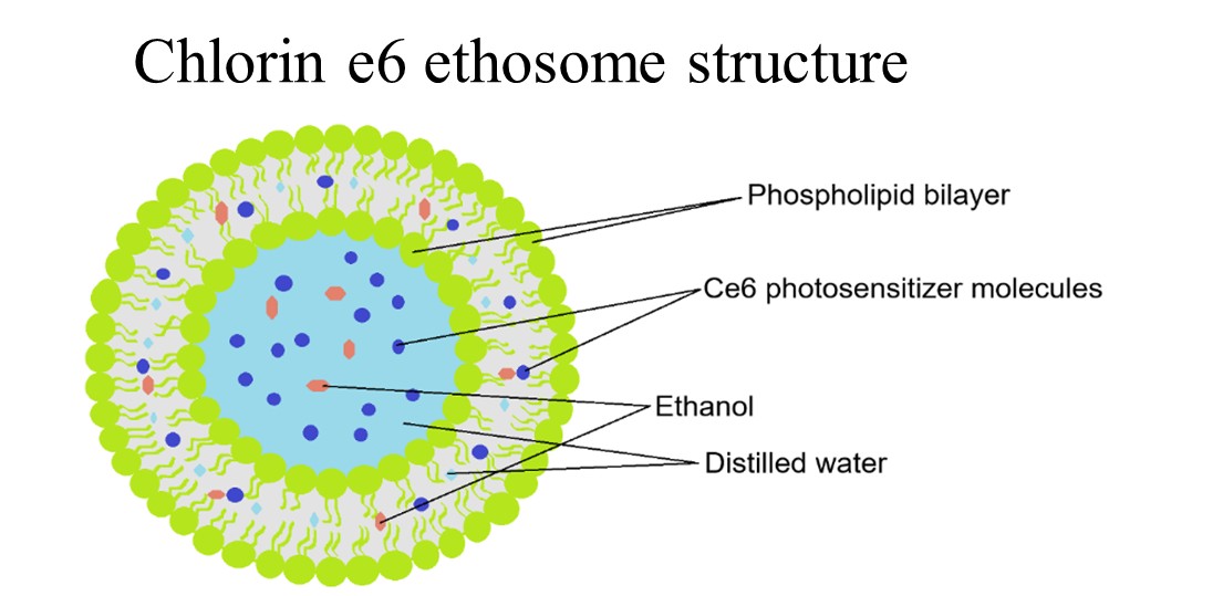
Chlorin e6-loaded ethosomes for photodynamic wound therapy
Limitations of the porcine model in post-total laryngectomy research

