News and Announcements
Volume 3 Issue 4 2022
Order by
Download Full Issue
Latest time
EDITORIAL
Celebrating the 2nd anniversary of Biomaterials Translational
Zhidao Xia,
Qian Wang
3 Download 1192 Views
VIEWPOINT
Skeletal interoception: an emerging area for musculoskeletal research
Zhidao Xia
6 Download 1251 Views
VIEWPOINT
Engineered exosomes for future gene-editing therapy
Haoyu Guo,
Xin Huang
11 Download 1423 Views
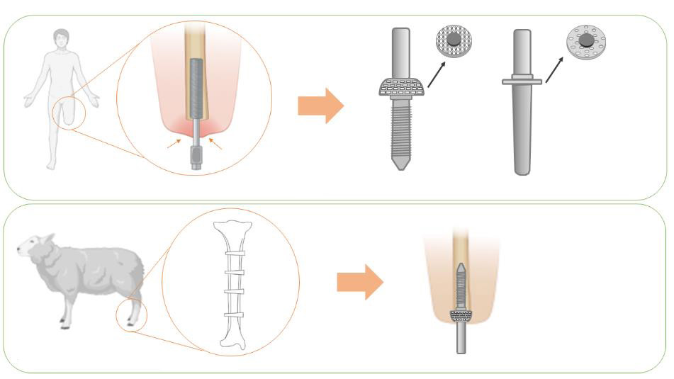
RESEARCH ARTICLE
Optimising soft tissue in-growth in vivo in additive layer manufactured osseointegrated transcutaneous implants
Elena Giusto,
Gordon Blunn,
Roberta Ferro de Godoy,
Chaozong Liu,
Catherine Pendegrass
5 Download 1314 Views
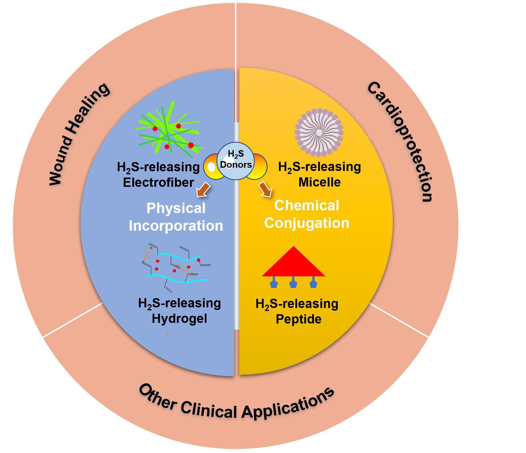
REVIEW
Recent development of hydrogen sulfide-releasing biomaterials as novel therapies:a narrative review
Jingyu Fan,
Elizabeth Pung,
Yuan Lin,
Qian Wang
30 Download 1729 Views
REVIEW
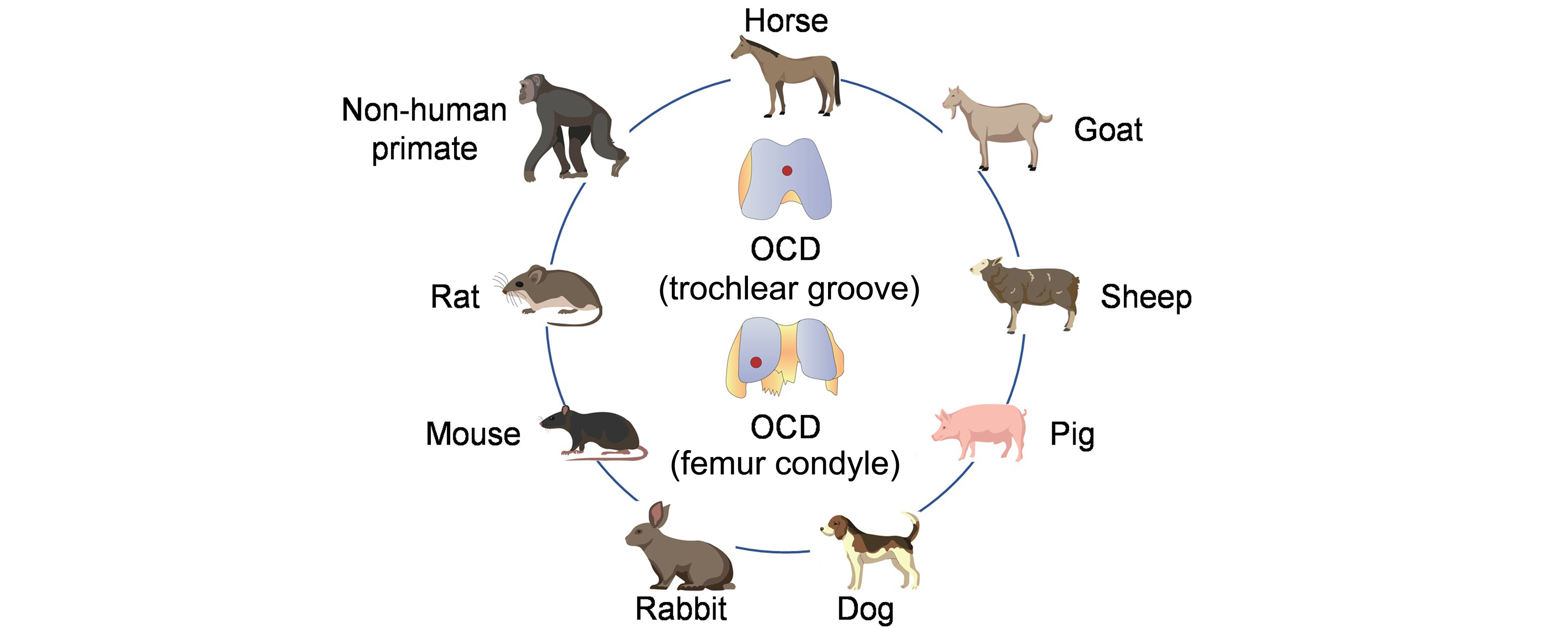
Osteoarthritis animal models for biomaterial-assisted osteochondral regeneration
Yi Wang,
Yangyang Chen,
Yulong Wei
34 Download 4472 Views
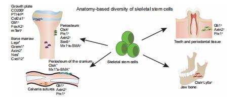
REVIEW
New perspective of skeletal stem cells
Guixin Yuan,
Zan Li,
Xixi Lin,
Na Li,
Ren Xu
11 Download 1564 Views