News and Announcements
Volume 5 Issue 3 2024
Order by
Download Full Issue
Latest time
EDITORIAL
Advances and challenges in hydrogel microspheres for biomedical applications
Yiting Lei,
Hélder A Santos,
Wenguo Cui
106 Download 3475 Views
REVIEW
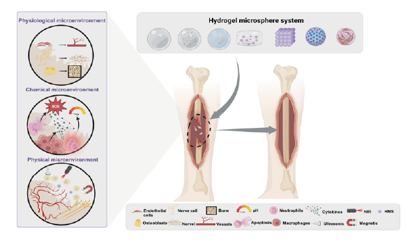
Hydrogel microspheres for bone regeneration through regulation of the regenerative microenvironment
Pengrui Zhang,
Qiwei Qin,
Xinna Cao,
Honglin Xiang, ... Yuling Li
202 Download 8933 Views
REVIEW
The use of hydrogel microspheres as cell and drug delivery carriers for bone, cartilage, and soft tissue regeneration
Chung-Hsun Lin,
Jesse R. Srioudom,
Wei Sun,
Malcolm Xing, ... Jian Yang
165 Download 6927 Views

REVIEW
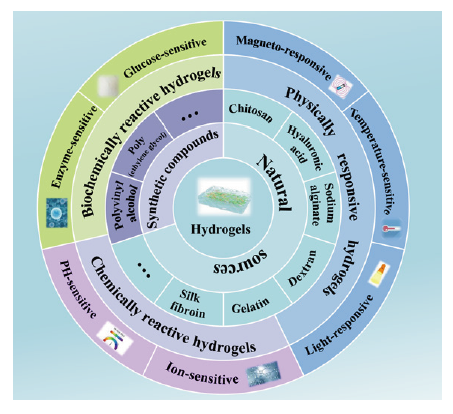
Stimuli-responsive hydrogels for bone tissue engineering
Congyang Xue,
Liping Chen,
Nan Wang,
Heng Chen, ... Xin Liu
98 Download 5399 Views
REVIEW
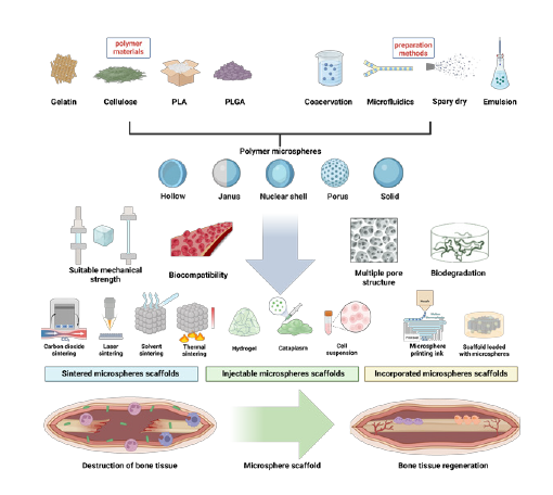
From the microspheres to scaffolds: advances in polymer microsphere scaffolds for bone regeneration applications
Shuhao Yang,
Haoming Wu,
Chao Peng,
Jian He, ... Xulin Hu
82 Download 4216 Views
RESEARCH ARTICLE
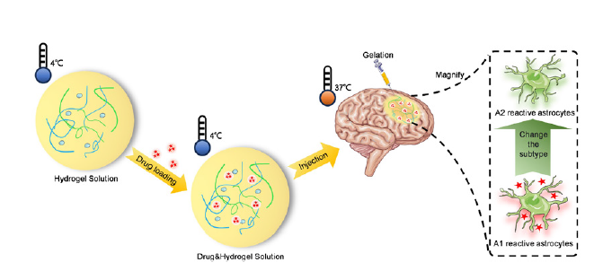
Injectable body temperature responsive hydrogel for encephalitis treatment via sustained release of nano-anti-inflammatory agents
Yuqi Gai,
Huaijuan Zhou,
Yingting Yang,
Jiatian Chen, ... Jinhua Li
154 Download 2705 Views
RESEARCH ARTICLE
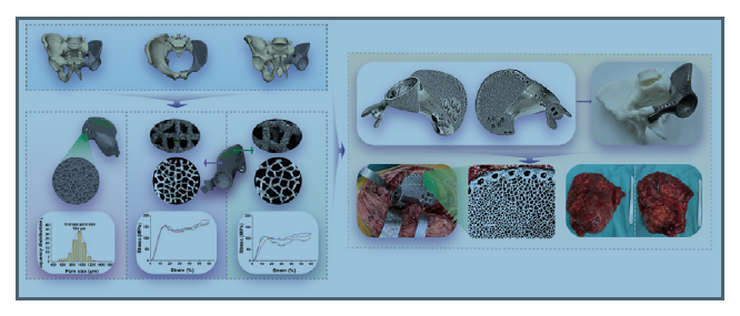
Design, characterisation, and clinical evaluation of a novel porous Ti-6Al-4V hemipelvic prosthesis based on Voronoi diagram
Zhuangzhuang Li,
Yi Luo,
Minxun Lu,
Yitian Wang, ... Chongqi Tu
82 Download 2207 Views
COMMENTARY
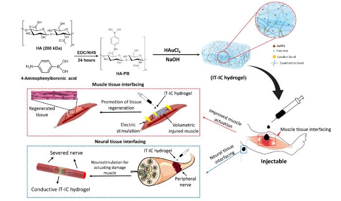
Bio-instructive hydrogel as an injectable tissue prosthesis for the repair and rehabilitation of impaired muscle
Muhammad Arif,
Tengbo Yu,
Qihui Zhou
27 Download 1364 Views
COMMENTARY
Targeting tumour-osteoclast interactions: a trigger-explosion system to combat bone metastasis
Ang Gao,
Huaiyu Wang
22 Download 1426 Views

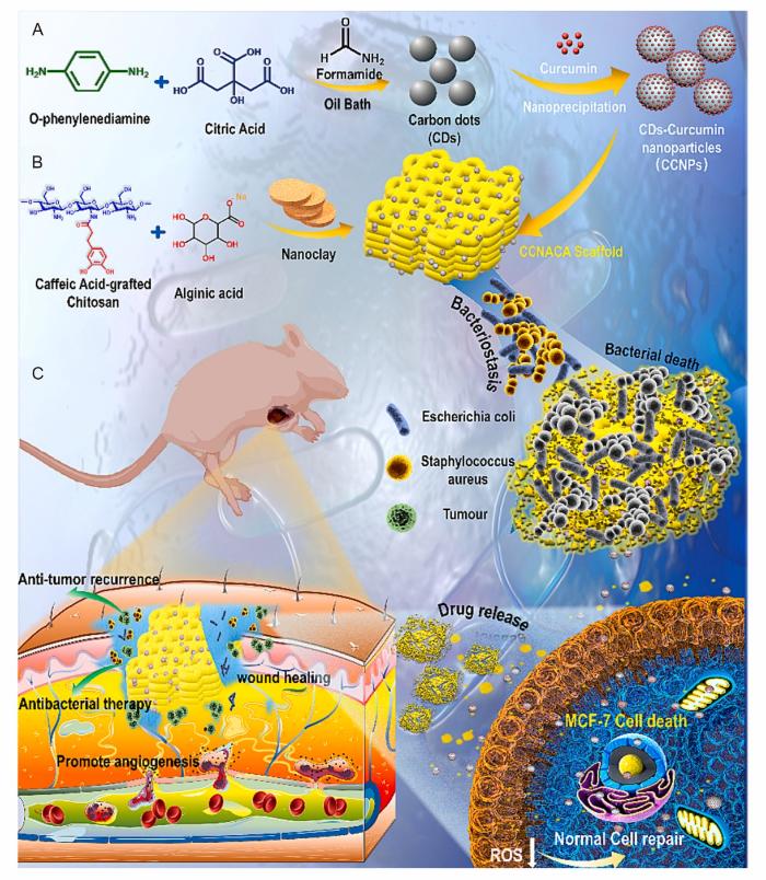
COMMENTARY
The potential of three-dimensional printed stents in post-operative treatment of breast cancer
Junjuan Fan,
Min Wang,
Xianwen Wang
21 Download 1407 Views